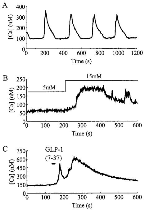
Fig. 1.
Representative examples of glucose and insulinotropic peptide-induced alterations of [Ca]i in β-cells. A, Slow oscillations of [Ca2+]i recorded when a β-cell was equilibrated in a steady state concentration (7.5 mM) of glucose. B, A β-cell was initially equilibrated in 5 mM glucose, then a stepwise increase in glucose concentration to 15 mM was applied by rapidly switching the contents of the bathing solution. C, A 30-sec pulse of 10 nM GLP-1-(7-37) was directly applied to a cell bathed in 5 mM glucose, using a puffer pipette (see Materials and Methods).

