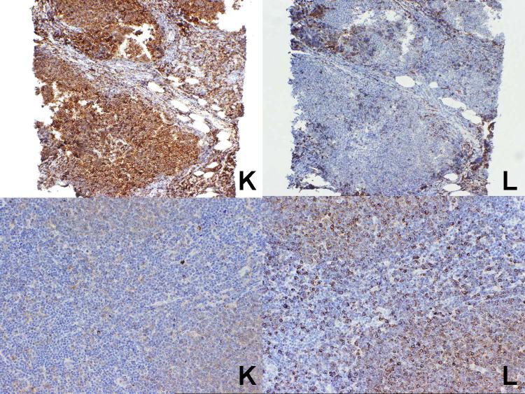
Figure 4.
Two cases of follicular lymphoma. At top is a case which was kappa light chain restricted, in agreement with the flow cytometry studies, while at the bottom is a lambda light chain restricted case, again in agreement with the flow cytometry studies. Note the weak staining of the follicles with kappa does not prevent assessment of the lower case as clearly showing lambda light chain restriction.

