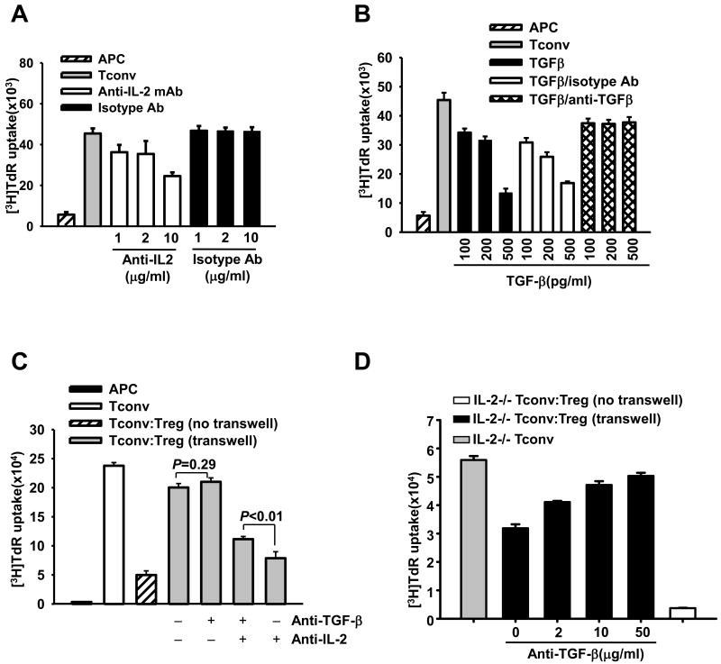
Figure 5. IL-2 deprivation creates prerequisite condition for TGF-β-mediated suppression by Tregs.
(A-B) Tconvs were stimulated by APCs and anti-CD3 mAb. In A, different amount of anti-IL-2 mAb or isotype control Ab was added into the cultures as indicated. In B, some cultures were added with TGF-β alone, or together with either anti-TGF-β mAb or isotype control Ab. (C-D) Tconvs isolated from wild type B6 (C) or IL-2−/− (D) mice were stimulated by B6 APCs and anti-CD3 mAb. Tconvs were cultured alone (Tconv group), or were co-cultured with Tregs from B6 mice either in the same chamber [Tconv:Treg (no transwell)] or in the different chambers [Tconv:Treg (transwell)] of a transwell. In C, cultures were treated with 10 μg/ml neutralizing anti-TGF-β mAb and/or 10 μg/ml anti-IL-2 mAb as indicated. In D, some cultures were treated with various concentration of anti-TGF-β mAb. Cell proliferation was assessed on day-3 by 3H-thymidine incorporation. Figures are representative of at least 3 independent experiments.

