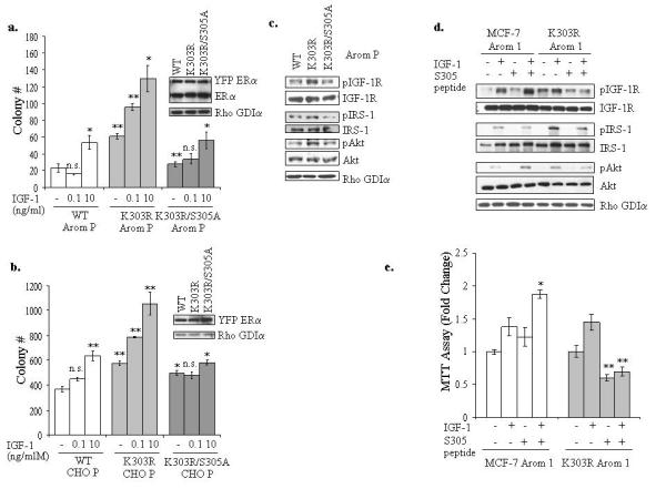
Figure 5.

Inhibition of serine 305 ERα phosphorylation blocked IGF-1R and mutant ERα cross-talk. (a, b) MCF-7 WT, K303R and K303R/S305A Arom P (a) and CHO WT, K303R and K303R/S305A P (b) pools of stably transfected and overexpressing cells were plated in soft agar and then treated with vehicle, IGF-1 0.1 or 10ng/ml for 14 days. The number of colonies >50μm were quantified and data are the mean colony number of three plates and representative of two independent experiments. Bars, SD. n.s.=nonsignificant, *P<0.01, **P<0.005. Equal expression of protein was determined by immunoblot with anti-ERα and Rho GDIα antibodies (right panel for a and b). (c) Cellular extracts from serum-deprived MCF-7 Arom WT, K303R K303R/S305A P pools of stably transfected and overexpressing cells were analyzed for phosphorylation (p) and expression of IGF-1R, IRS-1 and Akt by immunoblot analysis. Rho GDIα was used as a control for equal loading and transfer. (d) Cells were incubated with the S305 peptide (4μg/well) for 4h, and then treated with or without IGF-1 (10ng/ml) for 5min. Levels of phosphorylated (p) IGF-1R, IRS-1 and Akt, and total non-phosphorylated proteins were measured in cellular extracts by immunoblot analysis. Blots are representative of three separate experiments. Rho GDIα was used as a control for equal loading and transfer. (e) MTT growth assays in MCF-7 Arom 1 and K303R Arom 1 cells treated for 3 days with vehicle, IGF-1 (10ng/ml) and/or the S305 peptide (0.6μg/well). Cell proliferation is expressed as fold change relative to vehicle-treated cells. The data are representative of four independent experiments, each performed in triplicate. Columns, mean. Bars, SD. *P<0.005 compared to IGF-1 treatment in MCF-7 Arom 1-cells, **P<0.001 compared to vehicle and IGF-1 treatments in K303R Arom1-expressing cells.
