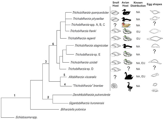
FIGURE 8.
Summary tree based on 28S depicting comparative features (hosts, distribution, and egg morphology) for North American and European avian schistosomes. Morphological features listed for well-supported nodes. 1– reduced sexual dimorphism, males and females flattened or thread-like, gynaecophoric canal absent or weakly developed or short (not extending to posterior), testes numerous; 2 – Absence of ventral sucker, absent or weakly developed oral sucker, uterus with numerous eggs, eggs ovoid; 3 – Well developed oral and ventral suckers, uterus usually with single egg, seminal vesicle between gynaecophoric canal and ventral sucker; 4 – cecal reunion at or anterior to seminal vesicle, >400 testes, gynaecophoric canal terminates well anterior to first testes; 5 – cecal reunion posterior to gynaecophoric canal, >400 testes, gynaecophoric canal terminates well anterior to first testes; 6 – Position of the cecal reunion overlaps the position of the seminal vesicle, gynaecophoric canal terminates at first testes, cercariae large with eyespots.
 Physidae,
  Lymnaeidae,
  Planorbidae,
  teal,
teal,  diving ducks,
  Anas americana
  most ducks,
  swans,
  geese. Eggs scaled to relative sizes.

