
Figure 3. Influence of clathrin-mediated endocytosis on Atg16L1-positive autophagosome precursors.
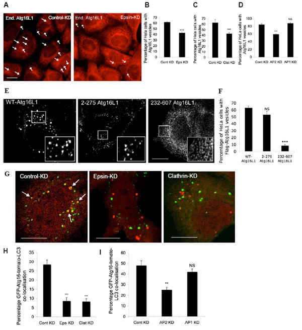
A, B, C, D. HeLa cells transfected with control, epsin 1, clathrin-heavy chain, AP2 or AP1 siRNA for 72 h were treated with Hanks balanced salt solution (to induce autophagy) for 6 h, after which they were fixed and immunostained for endogenous Atg16L1. The percentages of HeLa cells with Atg16L1 vesicles were quantitated. *** - p<0.0001. Scale bar – 10 μm. (Note that we used single siRNAs for all experiments and the effects were confirmed with two independent sequences for clathrin-heavy chain). n = 600 cells. E. F. HeLa cells transfected with Flag-tagged wild-type Atg16L1 or the Atg16L1 deletion mutants - 2-275 or 232-607 Atg16L1 for 24 h were fixed and imunostained with anti-Flag antibody and the proportion of cells with Flag-Atg16L1 vesicles were quantitated and represented in the graph. Representative images of Atg16L1 vesicles with the different mutant constructs is shown in the top panel. NS – not significant, *** - p<0.0001. n = 100 cells. G, H, I. HeLa cells transfected with control, epsin 1, clathrin-heavy chain, AP2 or AP1 siRNA, as above, were transfected with GFP-Atg16L1 and tomato-LC3 for a further 24 h, after which the cells were fixed. Percentage of GFP-Atg16L1 (green) that co-localised (yellow, marked by arrows) with tomato-LC3 vesicles (red) was quantitated as shown in the graph. n = 21 cells. *** - p<0.0001. Scale bar – 10 μm. All error bars in the graphs represent SEM.