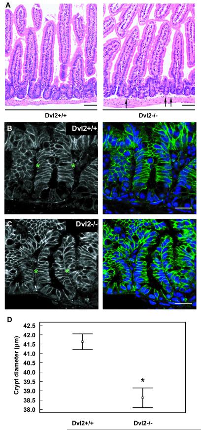
Fig. 4. Dvl2 mutants have reduced crypt diameters.
(A-C) Longitudinal sections through the small intestine of a 2 month-old Dvl2−/− and littermate controls, (A) after H&E staining; arrows point to individual narrow crypts in the mutant. Scale bars, 100 μm; (B, C) immunofluorescence images, revealing decreased crypt diameters in Dvl2−/− compared to littermate controls, as measured along apicobasal axis of intestinal crypt cells (between asterisks); blue, DAPI staining; green, α-β-catenin staining; scale bars, 25 μm. (D) Measurements of crypt diameters (n = 111) from sections as shown in (A) (4 mice per genotype); small squares indicate the mean, and lines standard error of the mean; *, statistical significance.

