
Fig. 2.
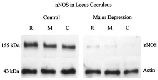
nNOS immunoreactivity in locus coeruleus (LC) from a single pair used in the analysis, representing three separate anatomical levels: rostral (R), middle (M) and caudal (C). Each well was loaded with 10 μg of total protein. The bottom panel (right and left) shows immunoreactive actin probed with anti-actin antibody on the same blot as a control for protein loading and transfer.