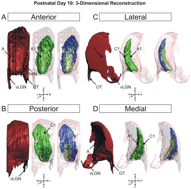
Figure 9.
3D reconstruction of the ferret dLGN at postnatal day 10 (P10). Perspectives facing the nucleus from the anterior pole (A), posterior pole (B), lateral aspect (C), and medial aspect (D) are shown. Contralateral projections are shown in red, ipsilateral projections are shown in green, overlapping projections are shown in blue for clarity. At this age, the major axis of the ipsilateral projections is oriented dorsoventrally. A, contralateral lamina; A1, ipsilateral lamina; C1, ipsilateral lamina; MIN, medial intralaminar nucleus; vLGN, ventral lateral geniculate nucleus; OT, optic tract.

