
Figure 4. Bivalent small molecule structure and library screening.
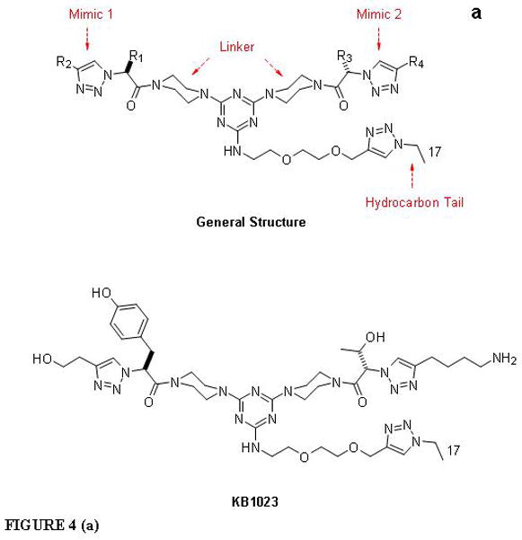

The general structure of the bivalent small molecule (a) includes two β-turn mimics for interaction with cell surface receptors, a hydrocarbon tail for insertion into BIV liposomal complexes, and a linker. The structure of our “hit” molecule, KB1023, is also shown. Our high-throughput luciferase assay (b) was used to screen for tumor endothelial cell-specific targeting ligands. At 7 days after co-culture, cells were harvested and seeded to 96-well plates at 2×10⁴ cells/well. On the same day, BIV-luciferase DNA:liposome complexes were prepared followed by coating of compounds at various compound:DNA ratios. The coated complexes were incubated at RT overnight. The following day, cells were transfected with 50 μL of serum free medium that contained 0.52 μL coated complexes. Transfection was ended by replacing the transfection medium with cell culture medium containing serum. At 24 h post-transfection, cells were lysed and the cell lysate was loaded to 96-well plates at 20 uL/well for luciferase assay using the Luminoskan plate reader.