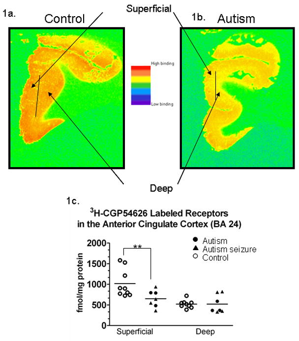
Figure 1.

Pseudocolored image of a control case (1a.) and an autism case (1b.) from the ACC off [3H]-sensitive hyperfilm. The images demonstrate the superficial (I–III) and deep (V–VI) layers that were sampled (1a.) In 1c, a graph demonstrating [3H] labeled GABAB receptor binding density in the anterior cingulate cortex. Each symbol represents the GABAB receptor density from an individual case (see key). There was a significant (**) reductions (p=0.018) in the density of receptors in the autism cases in the superficial layers. Note these are sample sections from individual cases, and although it appears that there is a difference in the deep layers in these cases, statistically there was no significant difference in binding density between autism and control cases.
