
Figure 6. Inhibitors of TLR7 decrease IFN-alpha secretion and CD80 expression in SV5-infected pDC.
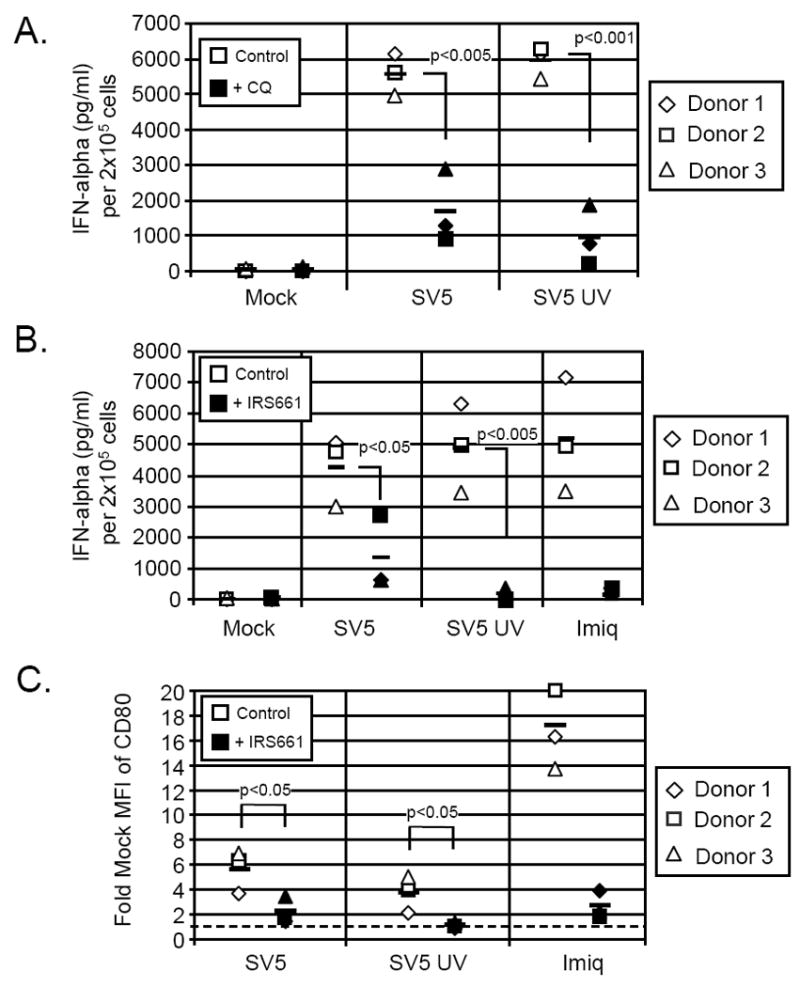
A) Effect of chloroquine. pDCs from three donors were mock treated (control samples) or pre-treated with 2.5 ug/ml of chloroquine (filled symbols). Cells were then mock infected or infected with live or UV-inactivated SV5 at an moi of 100. Twenty-four h pi supernatant was harvested and analyzed for levels of IFN-alpha by ELISA. B) Effect of TLR7 inhibitor. pDCs from three donors were pre-treated with 2 uM IRS661 (closed symbols) or were left untreated (open symbols) for 30 minutes. Cells were then mock infected or infected as described for panel A. Twenty-four h pi supernatant was analyzed for IFN-alpha by ELISA. C) Effect of TLR7 inhibitor on maturation markers. Cells were treated as described in panel B and at 24 h pi cells were stained for CD80 surface expression and analyzed by flow cytometry. Results are expressed as fold mock mean fluorescent intensity (MFI) of CD80 expression. The dashed line indicates the level of expression for mock infected cells set at one. The average of three donors for each condition is represented by the bar.