
Figure 1. Mammary duct epithelium of peripubertal C57BL/6 mice fed HFD has reduced outgrowth and terminal end bud formation.
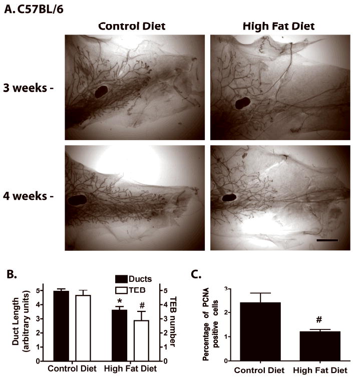
A. Whole mounts of representative mammary glands from peripubertal C57BL/6 mice fed CD or HFD for 3 or 4 weeks. Scale bar, 300 μm. B. Mammary duct length and TEB number in C57BL/6 mice fed CD or HFD for 4 weeks (n = 15). *, p < 0.001 or #, p < 0.05 when compared to CD fed C57BL/6 mice. C. Percentage of PCNA-positive epithelial cells in ducts, duct ends and TEB of C57BL/6 mice during estrus and fed CD or HFD (n = 3). #, p < 0.05 when compared to CD fed C57BL/6 mice.