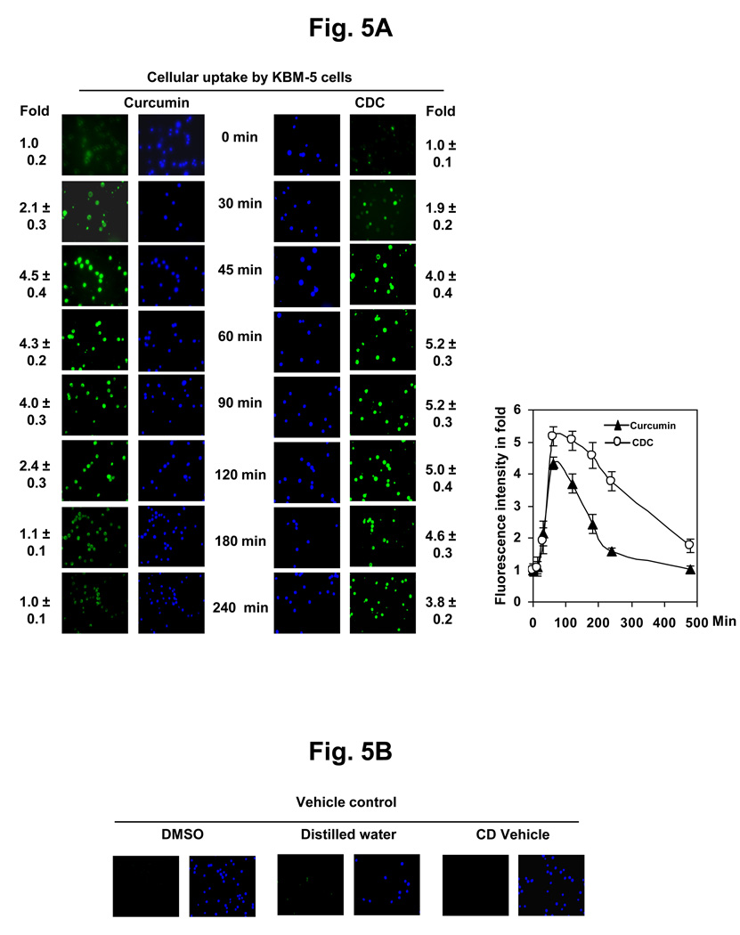
Figure 5.
(A) Cellular uptake of curcumin and CDC. KBM-5 cells (1 × 10⁶) were incubated with curcumin or CDC at concentrations of 10 µM. The cells were harvested at the indicated times, and the cellular uptake was determined by fluorescence microscopy as described in methods and blue color Hoechst’s staining showed cell viability. The results shown are representative of three independent experiments. (B) The control KBM-5 cells (2 × 10⁶) treated with DMSO and water. The results shown are representative of three independent experiments.

