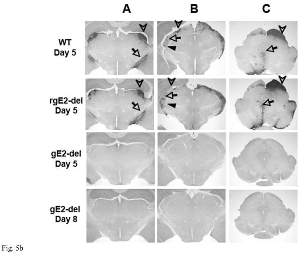
Figure 5.

A. The eye is shown at the bottom of the figure. Spread of viral antigens from the retina to the optic nerve and optic tract (OT) requires targeting of viral components from the neuron cell body into axons and anterograde axonal transport. Spread of antigen to the superior colliculus (SC), dorsal and ventral lateral geniculate nuclei (dLGN, vLGN) represents spread from pre-synaptic to post-synaptic neurons (anterograde spread). Spread of virus from the eye to the oculomotor nucleus (OMN) and Edinger-Westphal nucleus (EWN) requires spread from infected tissues in the eye to the innervating axons (epithelial to axon spread), retrograde axonal transport and spread from post-synaptic to pre-synaptic neurons (retrograde spread). B. Immunoperoxidase staining of mouse brains 5 or 8 dpi of the retina with 4 × 10⁵ PFU of WT, rgE2-del or gE2-del virus. The location of antigen is indicated by dark precipitate. Column A: HSV-2 antigen appears in the optic tract (requires targeting of viral antigens from the neuron cell body into axons and anterograde axonal transport, arrow) and the dLGN (requires anterograde spread, arrowhead) in WT and rgE2-del virus infected mice by 5 dpi but not in the brains of mice infected with gE2-del at 5 or 8 dpi. Column B: HSV-2 antigen is present in the dLGN (requires anterograde spread, arrowhead), the vLGN (anterograde spread, filled arrowhead) and the intergeniculate leaflet of the LGN (retrograde spread, arrow) in the brains of WT and rgE2-del virus infected mice but not in the brains of mice infected with gE2-del virus. Column C: HSV-2 antigen is detected in the SC (requires anterograde spread, arrowhead), the OMN and EWN (requires retrograde spread, arrow) of mice infected with WT or rgE2-del virus but not in the brains of mice infected with gE2-del virus. Results shown are representative of 3 mouse brains evaluated for each virus at each time point. Magnification is 20X.

