Fig. 1.

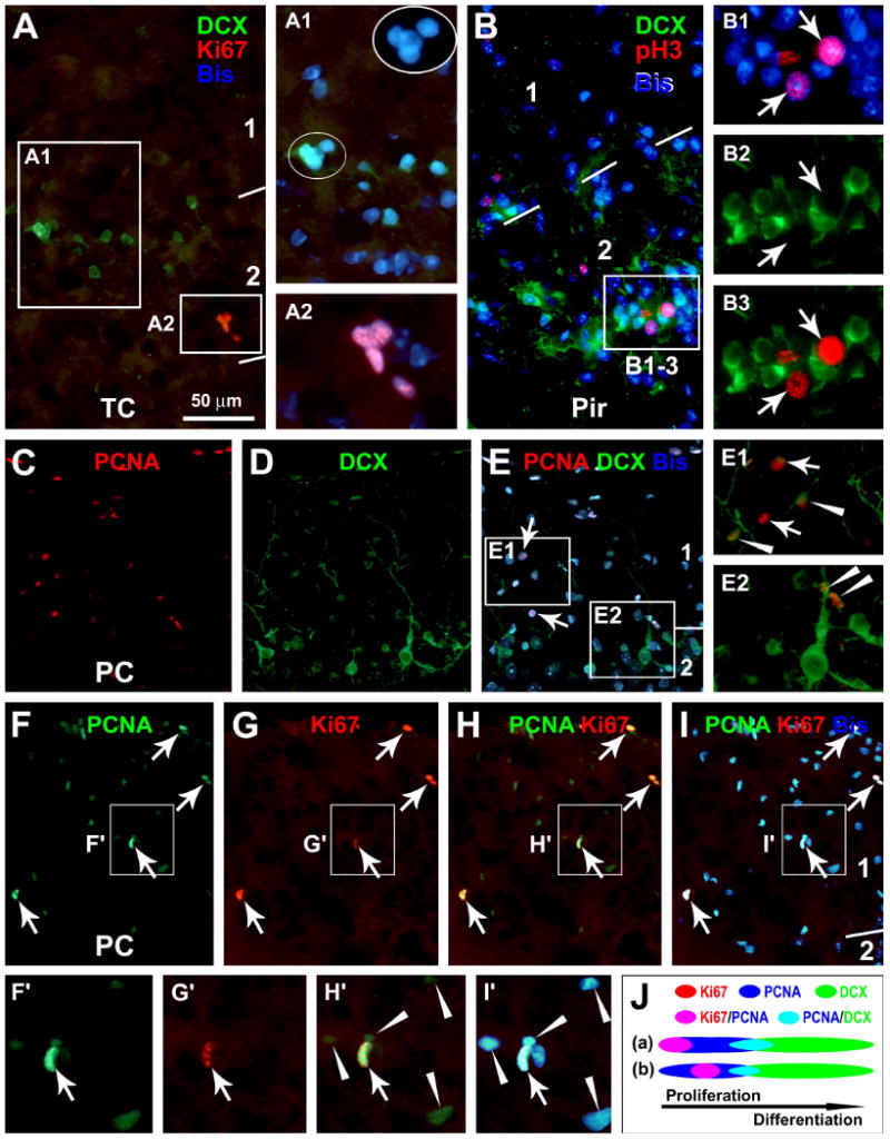
Representative images from 3 month-old guinea pigs showing the expression of endogenous proliferative markers, Ki67 (Ki67+), phosphorylated histone-3 (pH3+) and proliferation cell nuclear antigen (PCNA+), around layers I and II in relevance to doublecortin-expressing (DCX+) cells. Ki67+ (A-A2), pH3+ (B-B3) and PCNA+ (C-E2) nuclei occur discretely in layers I and II, including at the pial mater, with some labeled cells appear in pairs (C-I) or small clusters (A). Ki67+ and pH3+ cells may locate next to DCX+ cells in layer II (A, B), whereas PCNA co-localizes in some small DCX+ cells (C-E) (arrowheads). Among the PCNA+ nuclei, Ki67 co-expresses in those with heavy (arrows), but not those with weak (arrowheads), PCNA reactivity (F-I and F′-I′). The schematic draw (J) illustrates hypothetic temporal relationships for Ki67, pH3, PCNA and DCX expressions in layer I/II cells. Ki67/pH3 expression may begin at the same time (a) or somewhat later (b) but last shorter relative to PCNA during mitotic cell cycle. Arab numbers indicate cortical layers. Bisbenzimide (Bis) nuclear stain is shown in blue. TC: temporal cortex; Pir: piriform cortex; PC: parietal cortex. Scale bar in (A) = 50 μm applying to (B, E1, E2), equivalent to 25 μm for A1, A2, B1-B3, F′-I′; and 100 μm for (C-I).
