Fig. 2.

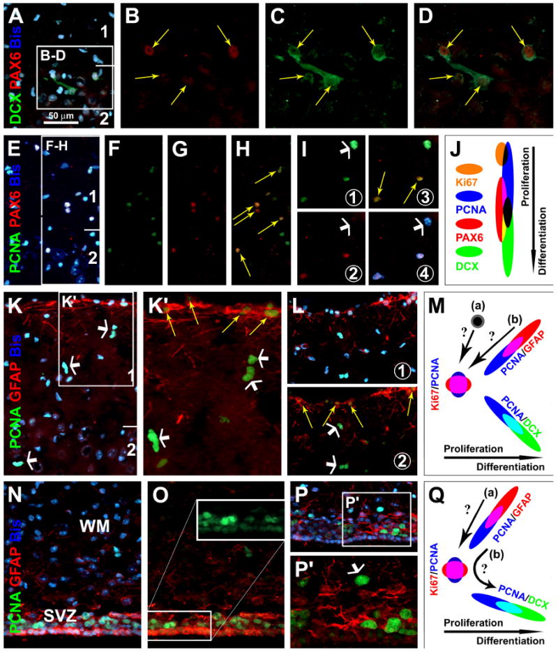
Partial colocalization of the Paired Box Protein Pax-6 (PAX6) with DCX (A-D) or PCNA (E-I) around layers I/II, and PCNA with glial fibrillary acidic protein (GFAP) in layers I (K-L) and the periventricular site (N-P) (images were from 6 month-old guinea pigs). PAX6 co-exists in DCX+ cells around layer II (yellow arrows), more distinct in small relative to large cells (A-D). PAX6 is partially colocalized among PCNA+ cells, in those with weak (yellow arrows) but not in those with heavy (white arrows) PCNA reactivity (E-I). GFAP+ cells around the pia may co-express weak PCNA (yellow arrows), whereas subpial PCNA nuclei (white arrows) lack GFAP colocalization (K, L). Around the lateral ventricle, PCNA and GFAP colocalize in ependymal layer cells (N-P). Schematic draw (J) depicts a hypothetic sequence of Ki67, PCNA, PAX and DCX expression among layers I/II cells, based on the partial colocalization patterns among these markers. Panel (M) hypothesizes two scenarios whereby the heavy-reactive PCNA cells around layer I may arise, from either specific proliferative layer I cells (a) or pia-associated GFAP+/PCNA+ cells (b). Panel (Q) depicts a potential astrocytic origin of immature neurons (PCNA+ → DCX+) around the subventricular zone (SVZ) (Ihrie and Alvarez-Buylla, 2008), either directly evolving from GFAP+/PCNA+ cells (b) or via a proliferative intermediate form (a). WM: white matter. Scale bar in (A) = 50 μm applying to (E-I, K, L-P), equivalent to 25 μm for B-D, K′ and P′.
