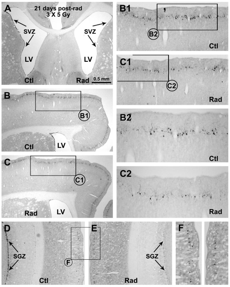
Fig. 5.

Representative images showing decline of DCX+ cells in the subventricular ventricular zone (A-C), subgranular granular zone (D, E) and around layer II of the neocortex (B-F) in the radiated (Rad) relative to the control (Ctl) sides 21 days after unilateral cranial X-ray irradiations (on day 1, 7 and 14). Framed areas in (B, C, D, E) are enlarged sequentially as indicated (B1, B2, C1, C2, F). Scale bar in (A) = 500 μm applying to (D, E), equivalent to 250 μm for (B1, C, F) and 125 μm for (B2, C2).
