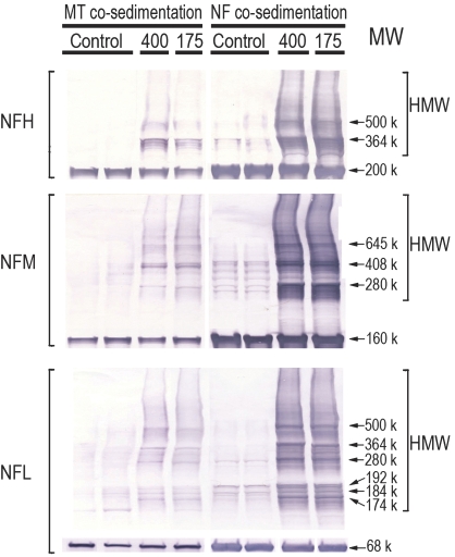
FIG. 4.
Representative immunoblots are presented for NF proteins (NFH, NFM, and NFH) in the final pellet of microtubule or NF preparations from control and HD-intoxicated (400 and 175 mg/kg/day dose rates) rats. Proteins were loaded on gels at 15 μg per lane. Immunoblots from HD samples illustrate the HMW derivatives associated with each NF subunit. The HMW laddering pattern for each subunit was comparable regardless of preparation method (i.e., microtubule vs. NF). In addition, similar HMW bands are clearly evident in control samples from microtubule or NF preparations.

