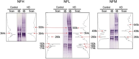
FIG. 6.
For each NF subunit, representative lanes from Figure 5B are presented from a control and an HD immunoblot (IB). The respective density scans (NIH ImageJ) for the HMW NF-immunopositive bands are provided on either side of the corresponding immunoblot. The immunoblots were calibrated to the HiMark prestained HMW protein standards.

