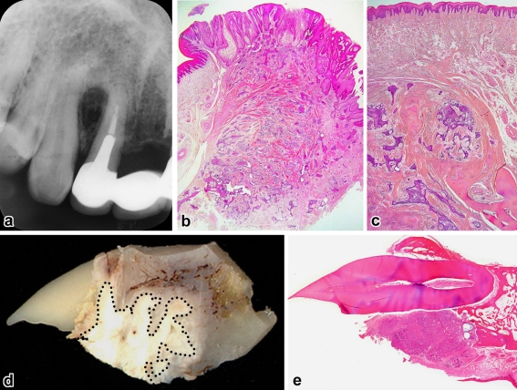
Fig. 2.
Cases 1 (a–c) and 4 (d and e). a Radiographic cupping of the interradicular bone, b gingival tumor showing scattered nests just near the excision edge (H&E, ×8), c recurrence in the bone without involving the surface epithelium (H&E, ×20), d decalcified gross specimen showing tumor location (dottedline), e surface ulceration and resorption of the palatal alveolar process (H&E, ×3)

