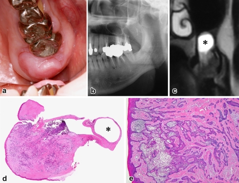
Fig. 4.
Case 5. a Exophytic nodular mass, b panoramic radiograph of bone destruction in the alveolar process, c T2-weighted MRI showing intraosseous tumor and mucosal cyst of the maxillary sinus (asterisk), d Scanning view of tumor and associated mucous retention cyst (asterisk) (H&E, ×3), e fusions of submucosal tumor with the gingival epithelium (H&E, ×40)

