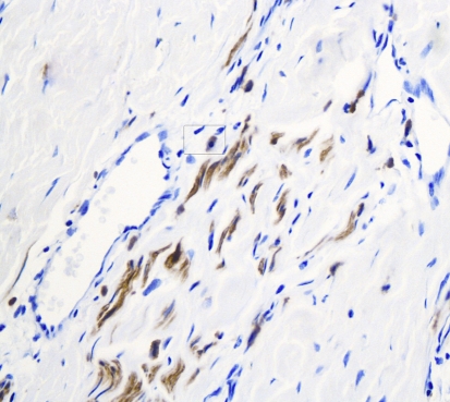
Fig. 3.
Immunohistochemical stain of S100 protein counterstained with hematoxylin, using horse radish peroxidase technique (40×). Note the positivity of thin spindled neural cells in short fascicles presented as brown/darkly stained cytoplasm. Note the presence of a mast cell in the rounded rectangle

