
Fig. 6.
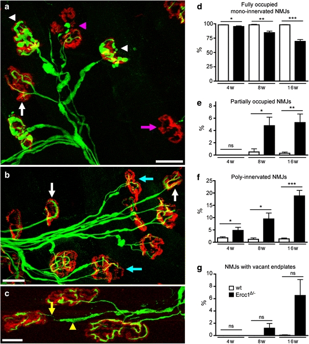
Synaptic pathology at the neuromuscular junction in Ercc1 Δ/− mice. a–c Confocal micrographs of neuromuscular junctions from lumbrical muscles of 16-week-old Ercc1 Δ/− mice labeled with rhodamine α-bungarotoxin to reveal post-synaptic motor endplates (red) and antibodies against 150 kDa neurofilaments to reveal pre-synaptic axons and motor nerve terminals (green). Examples of ‘normal’ (white arrows), denervated (pink arrow, a), partially denervated (pink arrowhead, a), poly-innervated (cyan arrows, b), and neuromuscular junctions are shown. Examples of motor nerve terminals with conspicuous neurofilament accumulation are indicated by the white arrowheads in a. Also note the presence of axon blebbing in c where the incoming axon is very thin (yellow arrow), but is interspersed by regions of neurofilament accumulations (yellow arrowhead). d–g Quantification of synaptic pathology in 4–16-week-old Ercc1 Δ/− mice compared to wild-type controls. *,**,***P < 0.05, 0.01, and 0.001, respectively, compared to wt mice of the same age (Kruskal–Wallis test with Dunn’s post-hoc test). Scale bars 40 μm (a, b) and 20 μm (c)