Figure 1.
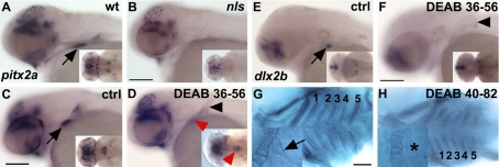
RA is required for pharyngeal tooth induction. A, B) pitx2a expression is absent in the ventral posterior pharynx in nls/aldh1a2 at 56 hpf (B) as compared with wild types (WT; A). C–H) Wild-type embryos treated with DEAB from 36 hpf (C–F) or 40 hpf (G, H) onward fixed at 56 hpf (C–F) or 82 hpf (G, H). In DEAB-treated embryos, pitx2a is faintly detected in the pharyngeal epithelium (D, red arrowhead) and is located in a group of cells at the midline (D, inset, red arrowhead). dlx2b expression is not detected in tooth buds in DEAB-treated embryos at 56 hpf (F). Alcian blue staining at 82 hpf of a control embryo (G) shows all branchial arches numbered from 1 to 5, including teeth (G, inset). In DEAB-treated embryos, all ceratobranchial arches are present (H), but teeth are absent. Asterisk marks the absence of tooth. Black arrowheads denote the presence of the pectoral fin that is present when DEAB is applied at late stage (later than 13 hpf; ref. 36). Scale bars = 100 μm.

