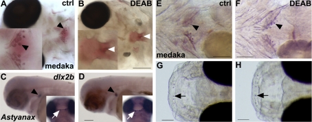
Figure 4.
RA is not required for oral and pharyngeal tooth induction in noncypriniform teleosts. A, B) Alizarin red staining of medaka embryos treated with DEAB during somitogenesis shows lack of pharyngeal teeth (white arrowhead in B) and the most posterior ceratobranchial arch (B), confirming the potency of DEAB in these species (ventral views). C, D) A. mexicanus control embryo (C) and DEAB-treated embryo from 30 hpf onward (D), showing dlx2b expression in oral (white arrow) and pharyngeal (black arrowhead) teeth (lateral views). E–H) Alizarin red staining of medaka embryos treated with DEAB from 3–7 dpf shows pharyngeal (F, arrowhead) and upper oral teeth (H, arrow; lateral views). Scale bars = 100 μm.

