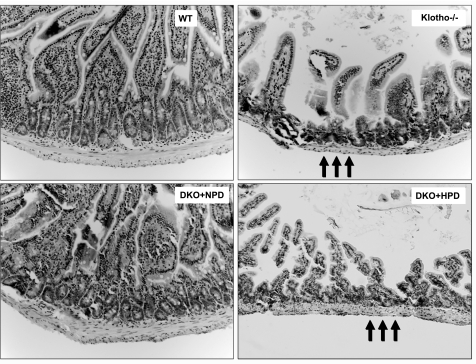
Figure 5.
Histological features of intestine. Hematoxylin and eosin-stained sections of intestines from WT, klotho−/−, DKO+NPD, and DKO+HPD mice. Compared with WT mice, there is marked atrophy of the intestinal wall (arrows) in klotho−/− mice. These focal atrophic changes of the intestine are improved in DKO+NPD mice and reappear in DKO+HPD mice, suggesting that reducing serum phosphate levels in the klotho−/− mice can help to restore intestinal anomalies (intestine view ×20).

