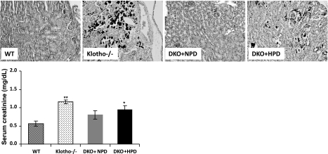
Figure 7.
Ectopic calcification of the kidney. Kidney sections prepared from WT, klotho−/−, DKO+NPD, and DKO+HPD mice, showing extensive calcification (black staining) in kidneys of hyperphosphatemic klotho−/− mice. Inactivation of NaPi2a in klotho−/− mice reduces this calcification in DKO+NPD fed mice. However, ectopic calcification reappears in DKO+HPD-fed mice, implicating phosphate toxicity in such ectopic biomineralization (von Kossa staining; ×20). The renal damage by calcification is also appearent from the serum creatinine levels of the mutant mice (bottom panel). Note that compared with the WT mice (n=4), serum creatinine level is significantly increased in the klotho−/− mice (n=4), and DKO+HPD (n=4). No such significant elevation is noted in DKO+NPD mice (n=4). *P < 0.05, **P < 0.01 vs. WT.

