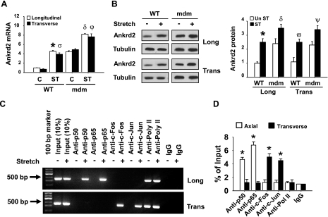
Figure 7.
Longitudinal and transverse directional stretches up-regulate Ankrd2 mRNA and protein expressions through the activation of Ankrd2 promoter in the mdm mouse diaphragm. Diaphragms of mdm mice were excised and stretched in ex vivo longitudinally or transversely to the myofibers for 15 min or maintained under unstretched conditions. A) Estimation of Ankrd2 mRNA by qPCR as described in Fig. 1A. Level of Ankrd2 mRNA is presented relative to that of GAPDH mRNA level (normalizer). B) Whole-cell lysates prepared from each diaphragm (85 μg/sample) were resolved by SDS-PAGE gel and immunoblotted with Ankrd2 or tubulin (loading control) antibody to show Ankrd2 and tubulin protein expressions. In another experiment, immediately after stretch, the diaphragms were cross-linked with formaldehyde and processed for ChIP assays as described in Fig. 2B. C) Agarose gel pictures show the binding activity of AP-1 or NF-κB on the Ankrd2 promoter. The fraction of chromatin used in the ChIP is shown (1% input). D) Equal amounts of DNA from the above samples were subjected to real-time qPCR assays to estimate Ankrd2 or GAPDH promoter DNA levels. Values represent the mean ± se of 3 independent experiments. *P < 0.05 for corresponding stretch and nonstretch samples. Greek letters on bars indicate significant difference to respective control. Gel pictures are representative of 3 independent experiments (n=3).

