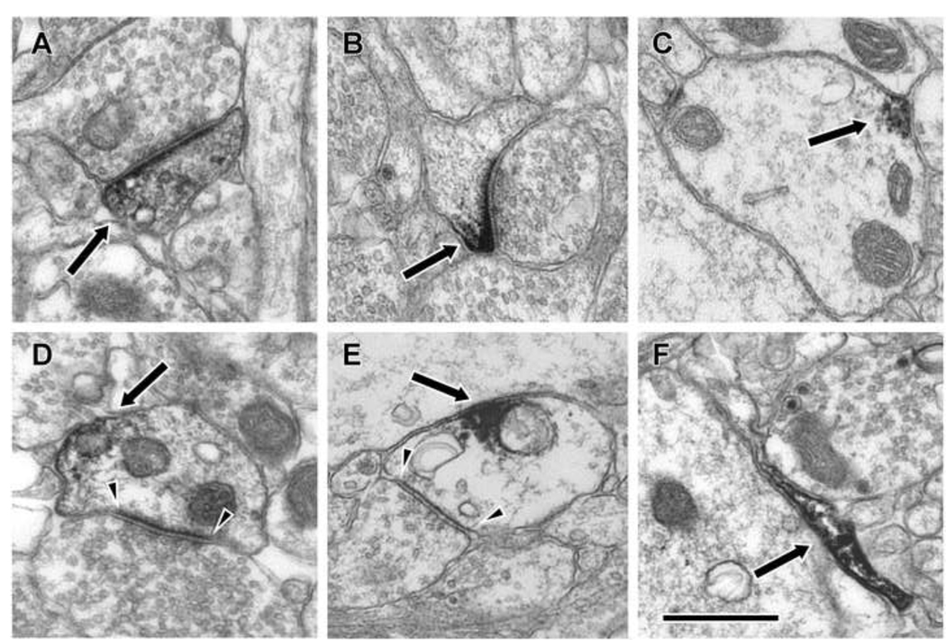
Figure 4.
Electron micrographs illustrating examples of D1R-IR in BLA neuropil. Diaminobenzidine label (arrows) was identified in spines for both D1 (A) and and D5 (B). Examples of dendrites containing D5-IR (C) and D1-IR (D, E) were also frequently identified. These labeled dendrites were sometimes observed to receive asymmetric (arrowheads, D) and symmetric (arrowheads, E) synaptic contacts. In addition to label in the dendritic arbors of neurons, label was also observed in glial processes as illustrated in F for a D1-IR process. Scale bar is 500 nm.

