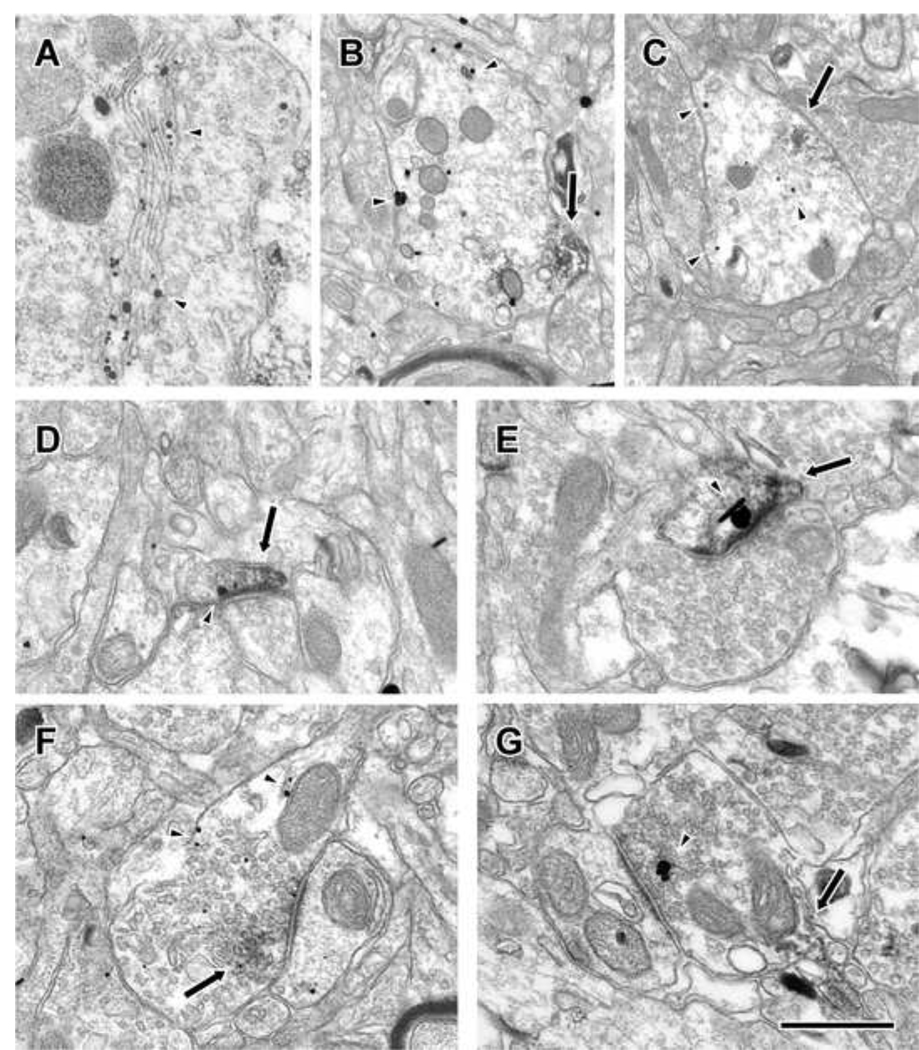
Figure 7.
Electron micrographs of double-label immunogold (arrowheads) and DAB (arrows) images of D1 and D5 in PFC. A: D1-gold label in the golgi apparatus of a cell soma. B, C: Double-labeled dendrites in which either D1 (B) or D5 (C) is labeled with gold. D, E: Double-labeled spines in which either D1 (D) or D5 (E) is labeled with gold. F, G: Double-labeled axon terminals in which either D1 (F) or D5 (G) is labeled with gold. Scale bar is 500 nm, except in B and C where it is 900 nm.

