
Figure 2.
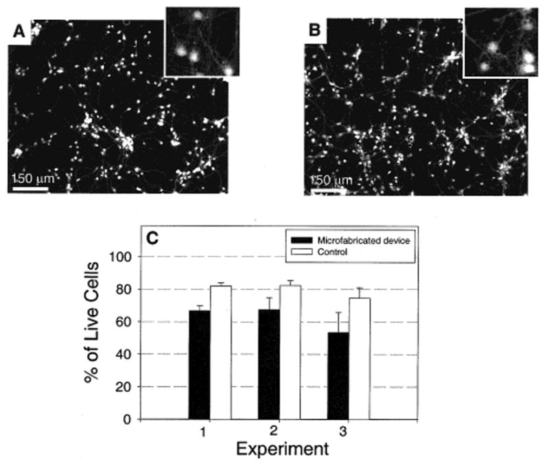
Viability of cultured primary neurons inside the microfabricated device versus tissue culture controls. (A) Fluorescence micrograph of neurons cultured for 7 days inside the microfabricated device. The PDMS top piece was sealed against a polylysine-coated tissue culture dish. Live cells were stained with a fluorescent probe, calcein AM (Molecular Probes). The box in the upper right-hand corner is a magnified section of the image showing the detailed morphology of the neurons. (B) Fluorescence micrograph of neurons cultured on a polylysine-coated tissue culture dish, without the PDMS device. Morphology typical of healthy neurons was evident in both microfabricated devices and controls. (C) Graph of cell viability inside the devices and controls after 7 days in culture (both used a polylysine-coated tissue culture dish as the substrate). The viability of cells cultured inside the microfabricated devices is slightly lower (10–20%) than that in controls, but they are generally comparable.