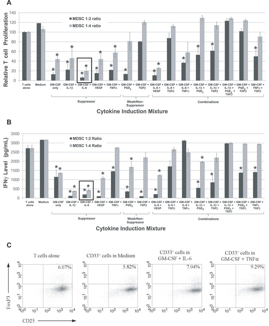
Figure 2. Cytokine-induced CD33+ MDSC demonstrate potent suppressive function.
PBMC from normal donors were cultured for one week in the presence of different cytokine mixtures. CD33+ cells were then isolated and tested for their ability to suppress the (A) proliferation and (B) INFγ production by autologous T cells at ratios of 1:2 and 1:4. CD33+ cells from cultures treated with GM-CSF and IL-6, Il-1β, VEGF, PGE2, or TNFα demonstrated suppressive function. For both graphs, mean is shown with SEM. Conditions with statistically significant decreases in mean T cell proliferation compared to stimulated T cells alone are indicated by an asterisk. (C) Treg expansion by cytokine-induced MDSC in Suppression Assays: the fraction of CD25+FoxP3+ T cells (CFSE-labeled) at the conclusion of a three day Suppression Assay with cytokine-induced CD33+ cells and fresh autologous T cells was analyzed by flow cytometry. Co-cultures with CD33+ cells induced by GM-CSF + IL-6 or GM-CSF + TNFα showed increases in CD25+FoxP3+ T cells relative to stimulated T cells cultured alone or with CD33+ cells from medium-only cultures (n=1).

