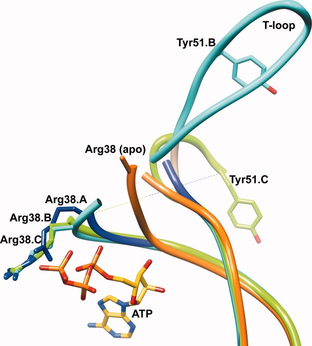
Figure 3.

Overlay of the T-loop regions of subunits A (blue), B (cyan), C (green) of the Mtb PII:ATP and Mtb apo PII (purple) structures, shown in a worm representation. The bound ATP molecule is shown in yellow stick representation. The relative positions of the Tyr51 residues on the T-loop of subunit B (cyan) and subunit C (green) of Mtb PII highlight the different orientations of the T-loop in both subunits. Arg38 of the Mtb apo PII protein (orange) is modeled as an Ala. Upon binding ATP, Arg38 of all three subunits of the Mtb PII:ATP structure (blue, cyan, and green) are stabilized and move relative to their position in the Mtb apo PII structure.
