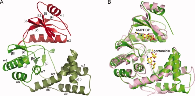
Figure 2.
(A) Ribbon representation of APH(2″)-IVa. The N-terminal domain is colored red and the C-terminal domain is in two shades of green representing the core subdomain (light green) and the helical subdomain (dark green). The numbering of the secondary structure elements is indicated. (B) Superposition of APH(2″)-IIa (pink) onto APH(2″)-IVa (green) based on all matching atoms. The location of the AMPPCP and gentamicin in APH(2″)-IIa are shown as ball-and-stick models (yellow). Figures 2–4, 5(A) and Supporting Information Figure S2 were prepared with the program PYMOL (http://pymol.sourceforge.net).

