
Figure 3.
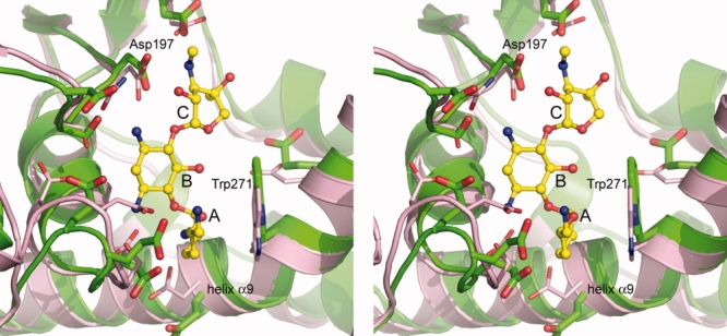
Stereoview of the superposition of APH(2″)-IIa (pink) onto APH(2″)-IVa (green) in the vicinity of the putative aminoglycoside substrate-binding site. The location of the bound gentamicin in APH(2″)-IIa is shown as a yellow ball-and-stick model, and the three rings have been labeled A, B, and C (see Fig. 1).