
Figure 4.
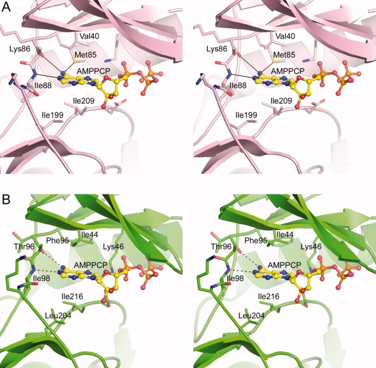
A: Stereoview of the nucleotide-binding site in APH(2″)-IIa18 showing the AMPPCP (yellow ball-and-stick model) and some of the residues which form the adenine-binding pocket. B: Stereoview of the putative nucleotide-binding site in APH(2″)-IVa showing the residues in equivalent positions to those in APH(2″)-IIa. The two potential hydrogen bonds that would anchor the adenine moiety are shown as dashed lines.