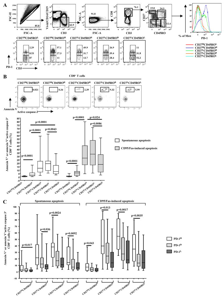
FIGURE 1.
The absolute expression of PD-1 is a primary indicator of ex vivo apoptosis of CD8+ T cells in HIV infection. A, The polychromatic flow cytometry gating scheme for identification of CD8+ T cell populations expressing low, dim, and high levels of PD-1 is shown. Histograms depict the PD-1 expression in naive and memory populations of CD8+ T cells from the same sample. Memory subsets identified by CD27 and CD45RO staining of total CD8+ T cells are also presented. B, Representative flow cytometry plots showing simultaneous measurement of annexin V binding and active caspase 3 levels in naive and memory CD8+ T cell from an HIV+ donor cultured for 12–14 h at 37°C (upper panel). Pooled data showing the percentage (%) of apoptotic naive and memory CD8+ T cells from HIV+ donors (n = 26) cultured in the absence or presence of anti-CD95/Fas Ab for 12–14 h (lower panel). C, Pooled data showing the percentage (%) of spontaneous and CD95/Fas-induced apoptosis in naive and memory CD8+ T cell compartments from HIV+ donors (n = 26) and in relation to expression of PD-1. Apoptosis sensitivity was evaluated based on annexin V binding or the simultaneous measurement of annexin V binding and active caspase 3 expression. Bars depict median values. p values were calculated using Mann-Whitney U test.

