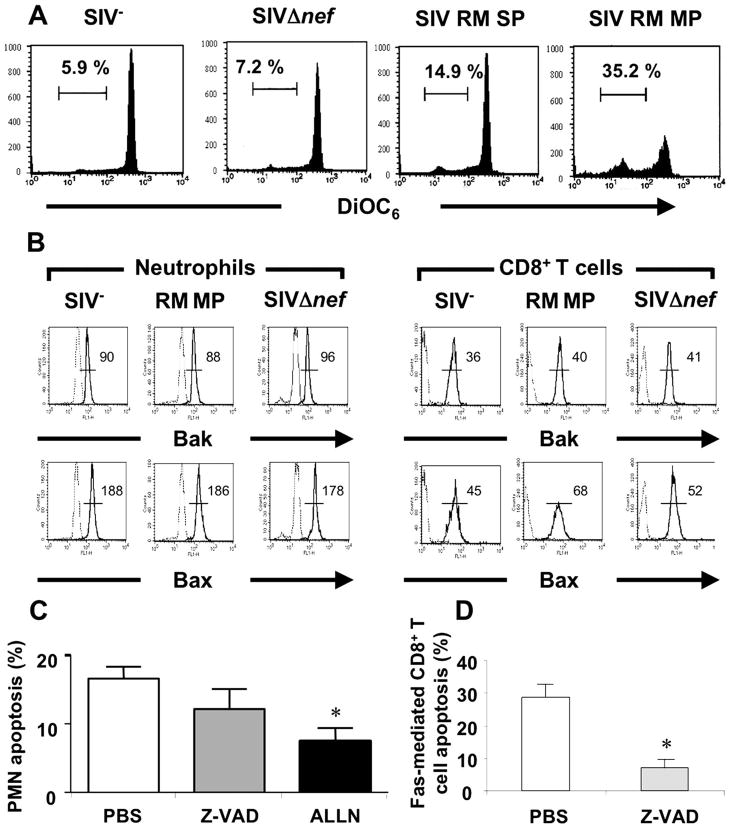
FIGURE 6.
PMN from SIV+ rhesus macaques display mitochondrial insult. A, Whole blood samples from healthy controls (SIV−), SIVΔnef and SIV+ RMs (slow progressors, RM SP, and moderate progressors, RM MP) on day 14 postinfection were incubated at 37°C for 4 h. Δψm loss was measured by flow cytometry using DiOC6 and expressed as the percentage of DiOC6low cells. One experiment representative of three. B, Bax and Bak staining of PMN and CD8+ T cells from healthy control (SIV−), SIVΔnef macaque, and RM moderate progressor (RM MP). One experiment representative of three is shown. C, Whole blood samples from SIV+ rhesus macaques on day 14 postinfection were incubated at 37°C for 4 h with PBS, z-VAD-fmk (a broad caspase inhibitor) (10 μM), or ALLN (a calpain inhibitor) (50 μM). Apoptosis was studied as described in the legend of Fig. 1. Values are means ± SEM (n = 6). D, Efficacy of z-VAD-fmk on apoptosis of macaques cells. CD8+ T cells from SIV+ RMs on day 14 postinfection were pretreated with PBS or z-VAD-fmk (10 μM) and then incubated in the presence of rhCD95L (200 ng/ml). T cell apoptosis was analyzed using annexin V+ staining. Values are means ± SEM (n = 3).*, p < 0.05, Significantly different from samples incubated with PBS.

