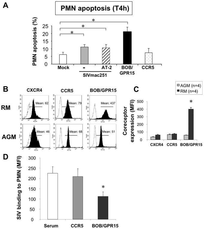
FIGURE 7.
SIV primes PMN for death. A, Whole blood samples from healthy controls (SIV−) were incubated for 4 h at 37°C in the absence (mock) or presence of 400 AID50 of the pathogenic SIVmac251 strain that has been either untreated (−) or treated with aldrithiol-2 (AT-2). Cells were also incubated with anti-BOB/GPR15 or anti-CCR5 Abs. Values are means ± SEM (n = 3). Purified sera were used as controls for chemokine receptors Abs; no difference in the percentage of dying cells was observed relative to the mock control (data not shown).*, p < 0.05, Significantly different from mock-incubated samples. B, Representative CXCR4, CCR5 and BOB/GPR15 expression on PMN from a healthy macaque and a healthy AGM, measured by flow cytometry. Dotted line histograms show isotype control stains. Filled histograms show specific Ab staining. C, Quantitative expression of CXCR4, CCR5, and BOB/GPR15 expression on PMN from healthy RMs (n = 4) and healthy AGMs (n = 4). Values are means ± SEM.*, p < 0.05, Significantly different from AGM. D, PMN from healthy controls (SIV−) were pretreated with purified serum, anti-CCR5 Ab, or anti-BOB/GPR15 Ab and then incubated in the presence of 400 AID50 of the pathogenic SIVmac251 strain. Binding at the cell surface was revealed by FITC-anti-gp120 (SIVmac251) Ab.*, p < 0.05, Significantly different from serum-incubated samples. MFI, Mean fluorescence intensity.

