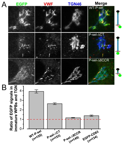
Fig. 7.
Immobile but not mobile P-selectin mutants become enriched in immature WPBs. (A) Fluorescence images of the Golgi region of fixed immunostained HUVECs expressing (top to bottom) wild-type P-selectin–EGFP, P-selectin-ΔCT–EGFP and P-selectin-Δ8CCR–EGFP; see molecule cartoons on the right. Cells were stained with fluorescent antibodies (from left to right) to EGFP (green in merged images), VWF (red in merged images) and TGN46 (blue in merged images). (B) The average ratios of EGFP immunofluorescence from perinuclear WPBs to that of adjacent membranes within the TGN (as described in Data analysis in the Materials and Methods) for these proteins and EGFP–CD63 are plotted. Bars show the s.e.m. Numbers of WPBs measured are shown in parentheses.

