Figure 5.

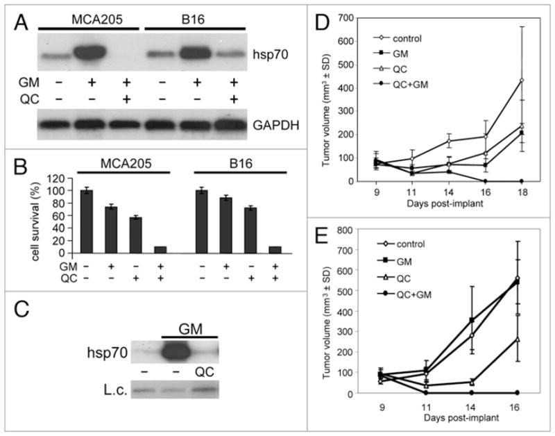
Effect of 17-DMAG and quinacrine on tumor cell growth in vitro and in vivo. (A) QC suppresses hsp70 synthesis in response to proteotoxic stress in MCA205 and B-16 cells. RNA from control cells and cells treated for 5 h with 1 μM 17-DMAG (GM) in the presence or absence of 20 μM QC was analyzed by northern hybridization with hsp70A1 and GAPDH probes. (B) Results of cell viability assay of MCA205 and B-16 cells treated for 4 h with 1 μM 17-DMAG (GM) in combination with 20 μM QC. Cells were harvested, diluted 1:50 and assayed for cell viability by methylene blue staining 72 h later. The data shown are the average of two experiments. (C) Intra-tumor injection of QC blocks 17-DMAG-induced hsp70 expression. C57BL/6 mice carrying MCA205 tumors were given a single intra-tumor injection of PBS (control), 25 μg 17-DMAG (GM), or 1.25 mg QC + 25 μg 17-DMAG. Five hours later, mice were sacrificed. RNA prepared from the tumors was analyzed by northern hybridization with a hsp70A1 specific probe. Hybridization with a GAPDH probe was used as an RNA loading control (L.c.). The data shown is from a single tumor for each treatment. These are representative data of two experiments. (D and E) C57BL/6 mice were injected with MCA205 (D) or B-16 (E) cells as described in Methods. Animals (n = 5 per group) began receiving intra-tumor drug injections on day 9 after tumor implant. The control group received PBS injections on days 9, 10, 11 and 12. Mice in group 2 (QC) were injected with 1.25 mg QC on days 9 and 10. Mice in group 3 (GM) received 25 μg 17-DMAG on days 9, 10, 11 and 12. Mice in group 4 (QC + GM) were injected with 1.25 mg QC + 25 μg 17-DMAG on days 9 and 10, and 25 μg 17-DMAG on days 11 and 12. Average tumor volume within each group is shown.
