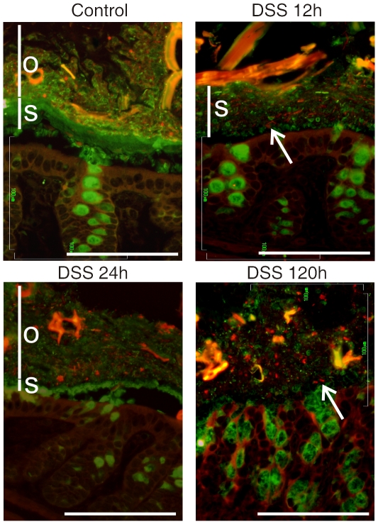
Figure 4. Localization of bacteria in the colon mucus of mice after DSS treatments for 12, 24 and 120 h or in nontreated control (No DSS).
Bacteria were stained by fluorescent in-situ hybridization using the general bacterial rRNA probe, EUB338 conjugated with Alexa 555 (red). The mucus was visualized by immunostaining of the same section with an anti-Muc2 specific antiserum (green). Penetration of bacteria through the inner firm and stratified (s) mucus layer was observed already after 12 h DSS administration. The inner stratified mucus layer is marked by s and the outer by o when any of the layers could be identified. Arrows point out bacteria within the inner mucus layer (12 h) or close to the epithelium in the absence of an inner mucus layer (120 h). Scale bars are 100 µm.

