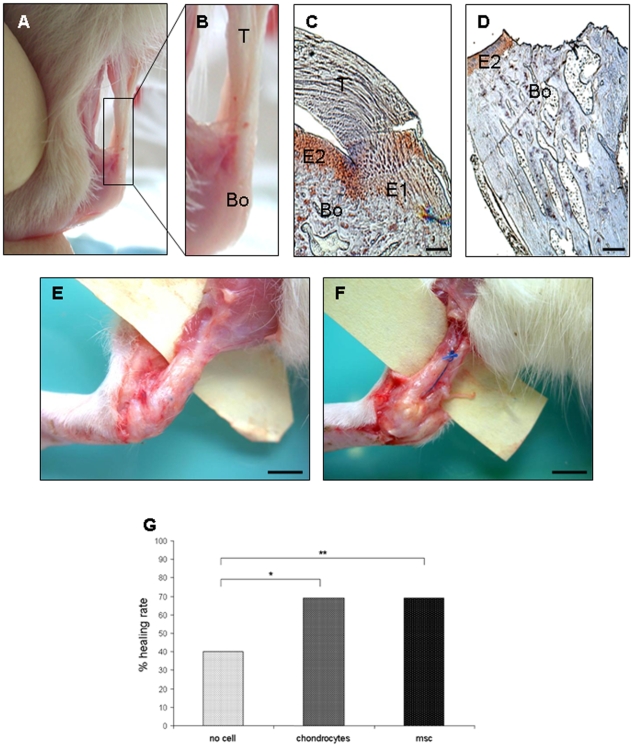
Figure 1. Enthesis structure and healing ratse after repair.
The enthesis is the area of bone-tendon junction. A) Wistar rat Achilles tendon-bone junction. B) Higher magnification of the delimited area shown in (A). T, tendon; Bo, bone. C and D) Type II collagen immunostaining of a native (C) and a destroyed enthesis (D). Native enthesis shows two positive areas of collagen II staining: E1, the insertion of the tendon (T) into the bone (Bo) and E2, the sliding zone of the tendon. Note the absence of the tendon and the E1 area just after destruction of the enthesis (D). (C and D, scale = 200μm). E and F) Healing failure after repair was evaluated by suture breakage (E) and a distance between the bone and the tendon of greater than 1 cm (F) (E and F, scale = 1 cm). G) Healing rate was evaluated at sacrifice for the three groups of rats and expressed as a percentage. * p<0.05 and ** p 0.01.

