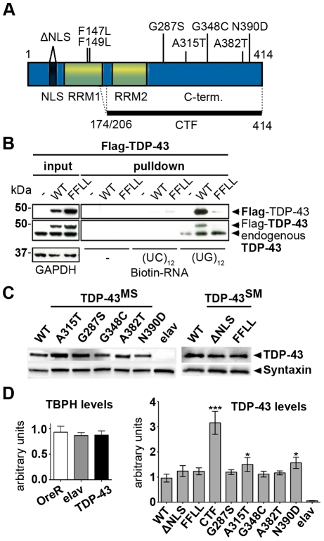
Figure 1. Analysis of different TDP-43 variants used for ectopic expression.
(A) Schematic overview of TDP-43 variants tested in vivo. Positions and nature of amino acid substitutions and size of two C-terminal fragments (CTFs) analyzed are indicated. Synthetic mutations (SM) were introduced to interfere with TDP-43 localization or function. Mutations in the nuclear localization signal (ΔNLS) were introduced to target TDP-43 to the cytoplasm and mutations F147L/F149L (FFLL) in the first RNA recognition motive (RRM1) were introduced to impair TDP-43 inherent RNA-binding capacity. (B) Loss of RNA-binding by TDP-43FFLL. Biotinylated oligonucleotides were incubated with lysates from either untransfected HEK293 cells or HEK293 cells transfected with FLAG-tagged TDP-43WT or TDP-43FFLL (left, input) followed by UV crosslinking and streptavidin-pulldown of Biotin-RNA (right, pulldown). Precipitates were separated, blotted and membranes probed with FLAG- (upper panel) and TDP-43-specific (lower panel) antibodies to assay co-precipitation of TDP-43. Note: Co-precipitation of endogenous TDP-43 with (UG)12-repeats in all lysates. Ectopic FLAG-TDP-43WT, but not FLAG-TDP-43FFLL co-precipitated with cognate UG repeats. GAPDH served as loading control for protein input. (C) Equalized pan-neural expression of φ-C31-inserted TDP-43 variants in adult Drosophilae. TDP-43 expression was visualized using an anti-human TDP-43 antibody. Syntaxin served as loading control. (D) Assessment of relative TBPH and TDP-43 expression levels. Left graph: Relative abundance of endogenous TBPH transcripts in relation to actin5C of wild type strain OregonR (OreR), the Gal4-driver line (elavC155::Gal4) and flies with pan-neural TDP-43 expression (elavC155::Gal4/Y;UAS::TDP-43WT/+). TBPH levels were not significantly different in analyzed genotypes. Right graph: mRNA abundance of TDP-43 in flies with pan-neural expression of the different TDP-43 variants normalized to actin5C and TBPH mRNA levels. Significant differences are indicated. *p<0.05; ***p<0.001. Head lysates of flies with pan-neural expression of TDP-43 were used for Western blot and qPCR analysis. elav::Gal4 flies without a TDP-43 transgene (elav) were used as negative control.

