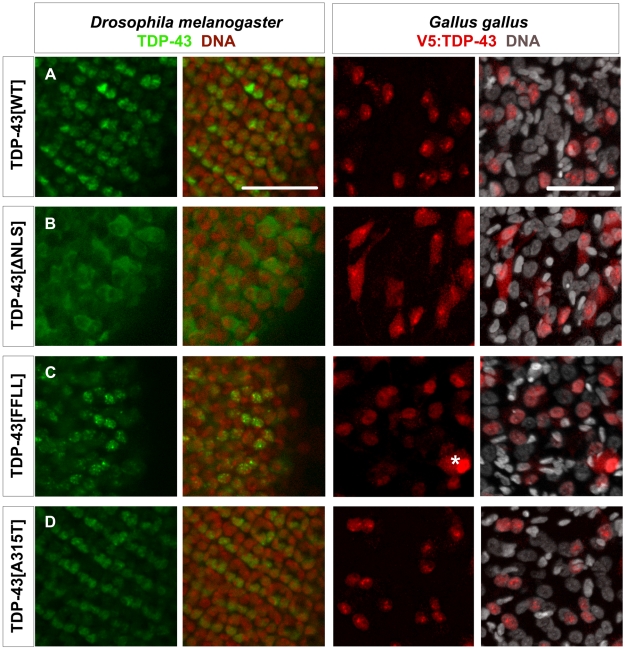
Figure 3. Localization of TDP-43 in Drosophila melanogaster and Gallus gallus.
Confocal sections of eye imaginal discs from Drosophila larvae (left panel) and motor neurons from Gallus (right panel) expressing indicated TDP-43 variants. To be able to discriminate between the two in vivo systems, ectopic TDP-43 in Drosophila is shown in green, whereas TDP-43 in Gallus is shown in red. Subcellular localization of the different TDP-43 variants was found to be identical between fly and chick. TDP-43WT (A) localized mainly to the nucleus, while TDP-43ΔNLS (B) was found predominantly in the cytoplasm. TDP-43FFLL (C) and TDP-43A315T (D) displayed a nuclear distribution. Only cells with very high expression levels of usually nuclear TDP-43 displayed a detectable cytoplasmic staining (example in case of TDP-43FFLL marked by asterisk). DNA was stained with Sytox® Orange (fly, red) or DAPI (chick, white). Scale bar indicates 50 µm. Neuronal expression was mediated by elav::Gal4 (flies) or Hb9::Cre (chick).

