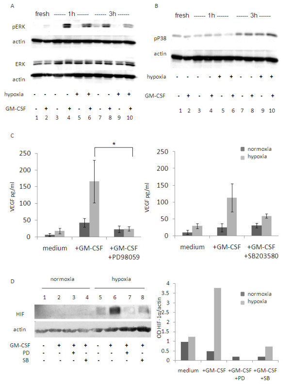
Figure 4.

Effect of GM-CSF signaling on hypoxia mediated HIF-1α and VEGF regulation. Eosinophils were incubated in normoxia/hypoxia in the absence or presence of GM-CSF. Western blot analysis was performed for phospho ERK1/2 and total ERK1/2 (A) or pP38 (B). The results are from one representative experiment out of three. (C) Eosinophils were cultured in medium with or without GM-CSF and with or without PD98059 (left panel; n = 5) or SB203580 (right panel; n = 3). MAPK inhibitors were added at the starting of the culture. VEGF levels were analyzed by ELISA. Data are the mean ± SEM of three experiments (* p ≤ 0.05). (D) Eosinophils were cultured for 3 h in medium alone (lanes 1 and 5), with GM-CSF alone (lanes 2 and 6) or with GM-CSF with PD98059 (lanes 3 and 7) or SB203580 (lanes 4 and 8). HIF-1α levels were analyzed by Western blot (left panel). Densitometry analysis is shown as a column chart (right panel). Data are representative of four experiments.
