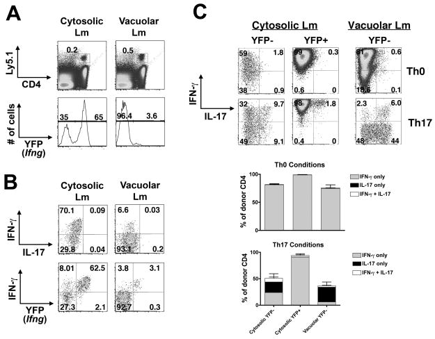
Figure 6. Previous Ifng expression marks Th1 committed cells.
A. P25 x Yeti CD4+ T cells were transferred into control B6 recipients 24 hours prior to infection with cytosolic or vacuolar Lm-Ag85B. Histograms represent percent donor cells that express Ifng (YFP+) directly ex vivo day 28 after infection. B. Representative FACS plots demonstrating IFN-γ and IL-17 production or YFP expression by cytosolic Lm or vacuolar Lm-primed CD4+ T cells after stimulation with cognate peptide directly ex vivo. C. Representative FACS plots (top) and composite bar graphs (bottom) demonstrating IFN-γ and IL-17 production by YFP− or YFP+ donor memory cells after culture in the indicated conditions for 5 days and restimulation with cognate peptide.

