
Figure 3. Casein Kinase I is involved in regulating Mdm2 stability.
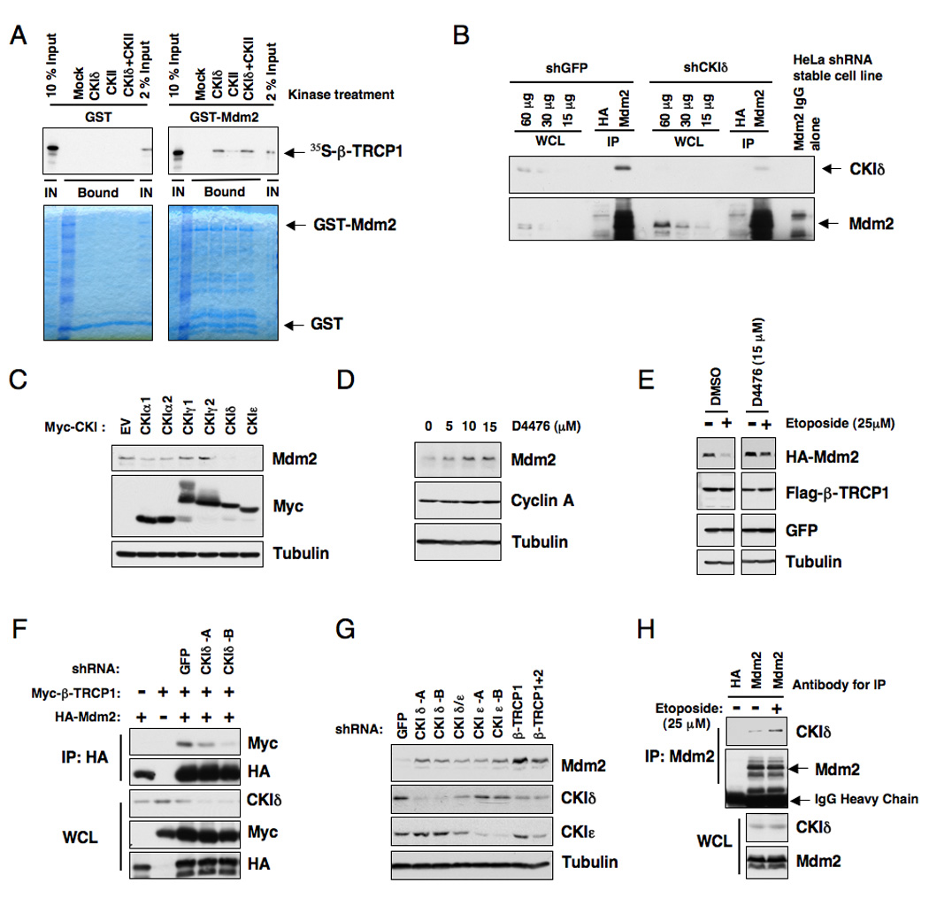
A. Autoradiograms showing recovery of 35S-labeled β-TRCP1 protein bound to GST-Mdm2 fusion proteins (GST protein as a negative control) incubated with the indicated kinase prior to the pull-down assays. IN, input (10% or 2% as indicated). 35S labeling was carried out by in vitro translation reaction with retic lysate.
B. Immunoblot analysis of whole cell lysates (WCL) and anti-Mdm2 immunoprecipitates (IP) derived from the indicated HeLa stable cell lines generated by infection with shGFP or shCKIδ lentiviral construct and subsequent selection with 1 µM puromycin to eliminate the non-infected cells. HA-agarose beads were used as a negative control for the immunoprecipitation procedure. Cells were treated with 10 µM MG132 overnight before harvesting.
C. Immunoblot analysis of whole cell lysates (WCL) derived from 293T cells transfected with various Myc-tagged CKI constructs to detect the changes in endogenous Mdm2 expression.
D. Immunoblot analysis of HeLa cells treated with the CKI inhibitor D4476 at the indicated concentrations for 12 hours.
E. HeLa cells were transiently transfected with the HA-Mdm2 plasmid together with Flag-β-TRCP1. Twenty-four hours post-transfection, cells were treated with the indicated CKI inhibitor for 4 hours and then incubated with 25 µM etoposide for an additional 1.5 hours. The whole cell lysates were recovered and immunoblots were performed with the indicated antibodies.
F. Immunoblot analysis of whole cell lysates (WCL) and immunoprecipitates (IP) derived from 293T cells transfected with HA-Mdm2 and Myc-tagged β-TRCP constructs. Where indicated, the shGFP or shCKIδ construct was included in the transfection. Twenty hours post-transfection, cells were treated with 10 µM MG132 overnight before harvesting.
G. Immunoblot analysis of HeLa cells transfected with the indicated shRNA constructs.
H. Immunoblot analysis of HeLa cell whole cell lysates (WCL) and anti-Mdm2 immunoprecipitates (IP). HA-agarose beads were used as a negative control for the IP. Cells were treated with 10 µM MG132 overnight before harvesting. Where indicated, cells were treated with 25 µM etoposide (or DMSO as control) for 30 minutes before harvesting. See also Figure S3.