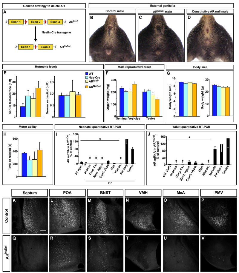
Figure 5. Targeted deletion of AR in the nervous system.
(A) Genetic strategy to delete AR in the nervous system.
(B–D) Adult external genitalia and milk line are masculinized in control and ARNsDel males, but not in constitutively null AR males.
(E) Similar levels of serum testosterone and estrogen in all males (n ≥ 12/genotype).
(F) Similar weight of testes and seminal vesicles in all males (n ≥ 7/genotype).
(G) Similar body length (snout to base of tail, n ≥ 4/genotype) and weight (n ≥ 12/genotype) in all males.
(H) No difference in time to fall from rotarod between control and ARNsDel males (n ≥ 4/genotype).
(I, J) Reduction in normalized AR mRNA in the brain of P1 (I), P7 (I), and adult (J) ARNsDel males shown as percent of AR mRNA levels in controls (Hypo., hypothalamus; Cing. Cx., cingulate cortex; Rost. Hypo., rostral hypothalamus; Caud. Hypo., caudal hypothalamus; Hippoc., hippocampus). Similar AR mRNA levels between ARNsDel and control males in other tissues. Mean ± SEM; ‡ mRNA < 0.5% of control, * p < 5×10⁻⁴, n = 4 for each genotype. (K–V) Fewer AR immunolabeled cells are visualized in coronal sections through septum, POA, BNST, VMH, MeA and ventral premamillary nucleus (PMV) in ARNsDel males compared to control males. Scale bar equals 100 μm.
See also Figure S3.

